Frame
Repair is a sequence, not “more myelin”
Oligodendrocyte precursor cells are recruited, then differentiate, then wrap axons, then compact a cholesterol-rich membrane. Failure can happen at any step.
B4 · @AndrewGilmerUS · Wednesday 9:00 PM EDT
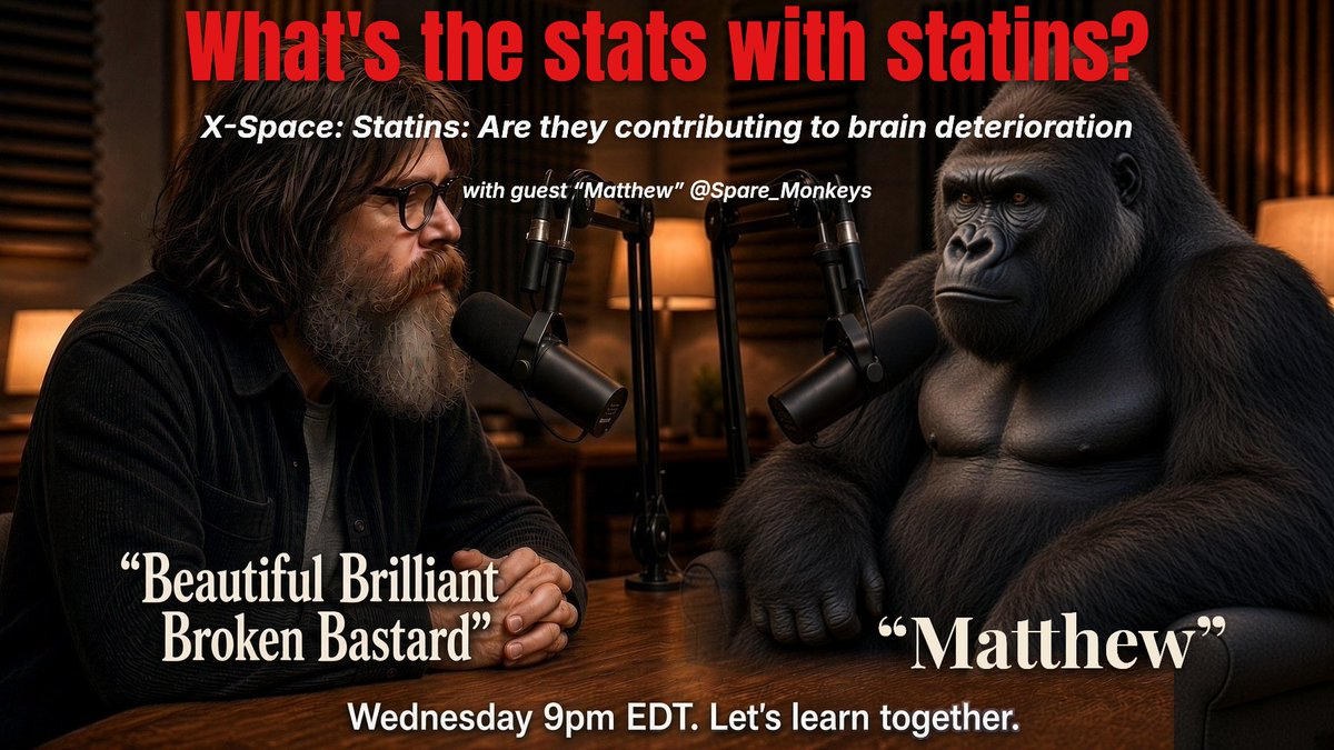
with guests: “Matthew” @Spare_Monkeys
Do you know someone who has been acting off past age 45? No promises. A conversation on whether statins may be contributing to brain deterioration.
Where statins came from, why people argue about them, and then the searchable notes. Tags are documented, plausible, or weak. This is not medical advice and not a diagnosis.
Remyelination needs three things at once: a closed barrier, a cholesterol budget, and a sleep and immune window. Statins cut the budget. DKA, chemo, and sleep apnea open the barrier. APOE4 and DHCR7 variants start you with a smaller budget. Anticholinergics and chronic THC scramble the signal to build. Age and myelin debris refuse the work order. Stack two or three and the shorts stay.
Oligodendrocyte precursor cells are recruited, then differentiate, then wrap axons, then compact a cholesterol-rich membrane. Failure can happen at any step.
Age after about 55, myelin debris and a dirty matrix, chronic inflammation or a leaky barrier, not enough cholesterol, and oxidative or mitochondrial failure in precursor cells.
The textbook case. Precursor cells often sit in the lesion and never finish differentiating. Age and disease duration both cut repair. Cortical lesions remyelinate better than white-matter lesions.
Antibody-driven astrocyte injury. Remyelination is secondary and often poor.
Primary myelin or oligodendrocyte failure. Hundreds of genes. Not all of them are “myelin genes.” Diagnosis is an MRI pattern plus sequencing, not a vibe.
Barrier breakdown, fibrinogen in the tissue, and scar-matrix deposition. Fibrinogen itself blocks precursor differentiation through BMP signaling.
Especially repeated or severe pediatric crises. Neutrophil enzymes chew tight junctions. Osmotic swings can produce osmotic demyelination. Oxidative stress in white matter is documented. The insult is the crisis and the barrier damage, not “statins causing DKA.”
Including functional deficiency when the serum number looks normal. Methionine synthase fails, so myelin-protein methylation fails. Classic subacute combined degeneration.
Can mimic the myeloneuropathy of B12 deficiency.
Thyroid hormone is a master differentiation signal for precursor cells. Low T3 or T4 slows the whole cascade.
LDL-receptor knockout animals show barrier leak, neuroinflammation, and early cognitive change. A large Norwegian human study found no extra dementia risk and no statin–dementia signal in FH. Treat as cardiovascular comorbidity, not a proven myelin-repair killer.
APOE4 holds cholesterol inside oligodendrocytes instead of sending it into myelin, and it weakens the barrier. Human statin data in E4 carriers is mixed to protective for dementia risk. That does not prove myelin is unharmed. Mechanism and outcome can diverge.
Glymphatic clearance and precursor proliferation both run during sleep. REM correlates with that proliferation. Chronic loss means thinner myelin and a repair window that never opens.
Chronic inflammation, barrier strain, and possible brain insulin resistance. Indirect, and common.
Weak as a remyelination claim. They travel with sleep loss, inflammation, and medications. Do not list them as primary myelin toxins.
HMG-CoA reductase inhibitors. Simvastatin, lovastatin, and atorvastatin are the lipophilic set. Brain cholesterol is made locally. Simvastatin impairs remyelination after chemical demyelination in animals. Turnover is slow, about 0.02 percent a day, so the risk framed here is chronic use plus a second insult, not a weekend of Lipitor. Cochrane 2016 and HOPE-3 did not show population-level cognitive harm. Muldoon 2004 is the small-trial decline. Frame it as repair-budget suppression, not “statins melt brains.”
Persistent precursor depletion, thinner myelin, and microglia-driven astrocyte activation. Wiping microglia in mice reversed the myelin and cognitive hit. Human brains after chemo show fewer Olig2-positive cells.
Kills precursor cells and mature oligodendrocytes at concentrations relevant to cerebrospinal fluid. Delayed, progressive myelin destruction in mice.
More toxic to neural progenitors and non-dividing oligodendrocytes than to many cancer lines. Cisplatin produces myelin decompaction.
CMF and FAC: cyclophosphamide plus methotrexate plus 5-FU, with or without doxorubicin. Decreased remyelination, stalled precursors, and myelin decompaction.
Infliximab especially, also adalimumab and etanercept. Soluble TNF through TNFR1 is inflammatory. Membrane TNF through TNFR2 is needed for remyelination. Non-selective blockade can trigger central or peripheral demyelination. Rare, real, and labeled.
Can unmask autoimmune demyelination.
Fingolimod or natalizumab withdrawal. Lymphocytes re-entering can produce explosive demyelination. That is iatrogenic rebound, not a myelin toxin while the person is still on the drug.
Diphenhydramine and hydroxyzine. They cross the barrier, carry a high anticholinergic burden, and are associated with brain atrophy and dementia risk at cumulative dose. Diphenhydramine’s sigma-1 antagonism disrupts neuronal cholesterol trafficking. The myelin link is indirect. Stacked with a statin, synthesis is down and trafficking is down.
Older bladder drugs such as oxybutynin, some tricyclic antidepressants, and some antipsychotics. Same signaling problem for oligodendrocytes that need cholinergic cues. Cognitive burden is the documented part. The myelin link is indirect.
Do not overclaim. Cognitive dulling, word-finding trouble, and paresthesias are well documented. The oligodendrocyte literature goes the other way: AMPA and kainate blockade, and protection of pre-myelinating oligodendrocytes in hypoxic-ischemic white-matter models. Flag it as a cognitive-burden drug, not a myelin-synthesis blocker.
Haloperidol can increase precursor proliferation but inhibit differentiation into myelinating cells. Atypical agents are mixed. Some muscarinic antagonists, including clemastine, benztropine, and quetiapine, promote differentiation in screens.
Clobetasol showed up as a remyelination promoter in drug screens. Chronic systemic steroids are a different story: metabolism, sleep, and mood.
Toxic leukoencephalopathy shows up in case literature. Not a clean remyelination-failure class.
Thirty diffusion-MRI studies, about 1,457 users: lower fractional anisotropy in the corpus callosum and superior longitudinal fasciculus. Earlier onset is worse. Oligodendrocytes express CB1 and CB2. Developmental THC can promote myelination in animals. Chronic adolescent or adult use disrupts it. Years of daily use have case-level toxic leukoencephalopathy. Edibles count: the liver makes 11-hydroxy-THC, which crosses the barrier more efficiently than smoked THC.
White-matter atrophy, thinner myelin, fewer mature oligodendrocytes. Fetal and third-trimester models show large drops in precursors and mature cells. Some fractional-anisotropy studies show no clear recovery with abstinence.
NNK thins myelin. Alcohol plus NNK is synergistic. Lifetime smoking associates with lower fractional anisotropy independent of other drugs.
Whippets or heavy dental exposure. Oxidizes B12 into a functional deficiency even when the serum level looks normal. Demyelination of the dorsal columns. Supplements do not protect if use continues.
Delays precursor differentiation, disrupts myelin ultrastructure, and hits barrier endothelium.
A neurotoxin, elevated in some MS brain samples, with common exposure from water, additives, and some pharmaceuticals. The mechanism is oxidative stress, barrier injury, and possible autoimmunity, not a single enzyme block. Chelation is a physician decision, not a home protocol.
Found in MS white matter more often than in controls in one autopsy series. Oligodendrocyte toxicity is plausible.
Crisis-level ketones are oxidative and hard on endothelium. Nutritional ketosis is not diabetic ketoacidosis. A brain held glucose-starved for a long time can lose metabolic flexibility. Slamming carbohydrates back onto a damaged barrier is a different insult than “butter is poison.” This is a plausible personal mechanism, not a published law that keto destroys myelin.
Excess dopamine is an oxidative load on oligodendrocytes. White-matter injury is documented in use disorders.
BPA, some pesticides, and persistent organics. Animal and developmental myelin effects exist. Human remyelination data is thin.
Do not list moderate coffee as a primary remyelination toxin. CBD-dominant products without heavy THC are not the same signal as chronic THC.
PLP1, X-linked. The prototype hypomyelinating leukodystrophy.
GJC2, also called connexin 47.
POLR3A, POLR3B, POLR1C. Also called 4H: hypomyelination, hypodontia, hypogonadism. A transcription-machinery disease.
Microtubules in the sheath.
Transcription factors for oligodendrocytes.
Hypomyelination with congenital cataract.
DHCR7, the last step of cholesterol synthesis. Malformations, intellectual disability, and autism in a large fraction. In SLOS, Cochrane found insufficient statin benefit and a signal for more adverse reactions. Heterozygous or hypomorphic sterol-gene variants may affect about 3 percent of people and raise vulnerability to any sterol-blocking drug.
Desmosterolosis (DHCR24), lathosterolosis (SC5D), and CHILD or CDPX2 (NSDHL, EBP). Same pathway, different enzymes.
Common and semidominant. Cholesterol trapped in oligodendrocytes, hypomyelination, and a leaky barrier. Not a leukodystrophy. It is the population-level genetic hit.
Metachromatic leukodystrophy (ARSA), Krabbe (GALC), Canavan (ASPA), X-linked adrenoleukodystrophy (ABCD1), Zellweger and other PEX genes, and Niemann–Pick C, where cholesterol trafficking fails and hypomyelination is secondary.
EIF2B1 through EIF2B5. Translation stress. White matter melts under fever or other stress.
MLC1.
An interferonopathy. White matter plus inflammation.
Peripheral myelin, listed because many chemo agents that hit central myelin are also toxic in CMT, including vincristine and taxanes. Different compartment, same vulnerability logic.
Remyelination is best under 55 and in the first decade of an MS-like injury. Aged precursor cells ignore differentiation cues.
Contains MAG, Nogo-A, and OMgp. Blocks maturation until microglia clear it.
Hyaluronan, CSPGs, and fibrinogen from a leaky barrier.
LINGO-1, Notch, Wnt and beta-catenin, and semaphorin 3A. This is why anti-LINGO trials existed.
About 4.5 days of deprivation thinned myelin roughly 8 percent in mice. Thirty-two hours of recovery did not restore thickness. Sleep-loss oligodendrocytes show endoplasmic-reticulum stress and cholesterol-transport failure. Boosting cholesterol transport into the sheath prevented the conduction and behavior hit in a 2026 PNAS paper. That is the cleanest mechanistic rhyme with statins in this list.
Precursors are more oxidatively fragile than mature oligodendrocytes. Reactive oxygen species upregulate Id2 and Id4 and block differentiation. Low glutathione sits in the same pile.
Stroke, DKA, chemo, APOE4, inflammation, sleep loss. Opening the gate is one injury. Everything else keeps it from closing.
No single item has to “cause it.” Chemo, a long statin course, a cognitive-burden drug, lost REM, and metabolic inflexibility can cut the repair budget from several directions at once. That is the comorbidity thesis, not a courtroom autopsy.
Do not say statins have a dedicated Department of Justice myelin case. This search did not find one. Nearby cases, on the money cards, are about promotion, delayed results, or generic delay. None of them is a myelin case.
Do not say it is. It isn’t, on this record.
Do not say Topamax strips myelin the way methotrexate does. Current evidence does not say that.
Different dose, different acidity, different outcome. Do not equate them.
Some E4 data goes the other way on dementia incidence. Do not state it as proven injury.